Hydrosalpinx: Examinations that Can Be Taken
Hydrosalpinx refers to a disease caused by the adhesion of the cervical tubes of women under the stimulation of some inflammation, which causes the tube wall to be too narrow. Once hydrosalpinx is not conducive to the process of fertilized eggs, it can have a great impact on pregnancy. So what examinations should hydrosalpinx patients have?

Doctor's physical examination
The cystic mass can be touchedon one or both sides of the pelvis. The movement is limited, and there may be a pain when pressing.
Laboratory examination
Smear examination of vaginal or cervical secretion: the doctor will take a small amount of secretion from the patient's vagina or cervix with a cotton swab, make a section, and then observe it under the microscope to determine whether there is pathogen infection.
Imaging examination
1. Ultrasonic examination
Ultrasound is the most commonly used non-invasive examination method. The first choice for asexual women is transabdominal ultrasound or transrectal ultrasound. In addition, the first choice for women without special requirements is transvaginal ultrasound. Generally, 3-7 days after menstruation, the bladder should be emptied before the examination.
The diameter of the fallopian tube in mild hydrocephalus < one point five Cm, medium ponding diameter One point five ~ 3cm, the diameter of serious water accumulation is more than 3cm.
2. Hysterosalpingography (HSG)
It has the characteristics of simplicity and intuitiveness. According to the diffusion of contrast medium, the examination indicates whether the fallopian tube is unobstructed and the information of pelvic disease. Generally, the examination should be carried out 3-7 days after menstruation is over. Patients with vaginitis need to be cured before HSG to avoid the spread of inflammation to the pelvic cavity.

HSG can help us to know whether the cervical tube is abnormal, whether the uterine cavity is abnormal, whether the length and shape of the fallopian tube are abnormal, and it can locate the hydrosalpinx.
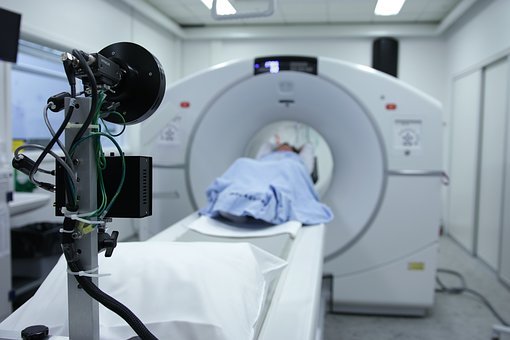
3. CT
CT has a high spatial resolution, which can provide more imaging data for the clinical diagnosis of hydrosalpinx.
4. Magnetic resonance (MRI) examination
This examination not only has the advantages of the high resolution of soft tissue and multi plain film imaging but also can better show the tubular structure of uterine horn and the situation in the pelvic cavity, which is conducive to the identification of salpingostomy and hydrosalpinx and can reduce the difficulty of diagnosis.
Special examination
Hysteroscopy is the gold standard for the diagnosis of hydrosalpinx. Laparoscopy can directly see the shape of the uterus and fallopian tube, the position of hydrosalpinx, the diameter of hydrosalpinx, and the surrounding pelvic conditions. Hysteroscopy can directly look at the cervix and uterine cavity for lesions, deformities and abnormalities of bilateral fallopian tube orifices.
You may also be interested in:
Diet for Blocked Fallopian Tubes due to Inflammation
previous pageWhich One Is Better, Natural Pregnancy Or IVF For Hydrosalpinx?
next page
You may also be interested in
- Hydrosalpinx and IVF: What Are the Chances of Successful Embryo Implantation?
- Is Hysterosalpingography Harmful to the Body?
- Can Hydrosalpinx Be Thoroughly Drained by Aspiration Surgery?
- Pay Special Attention to These Hydrosalpinx Symptoms!
- Hydrosalpinx: Frequent Checks and Excessive Anxiety Affect Pregnancy!
Testimonials
- Adenomyosis with Ureaplasma Urealyticum Cured by Fuyan Pill
- Tubal blockage with hydrosalpinx can be cured by TCM shortly
- Fuyan Pill Helps A woman with Adenomyosis Get Pregnant
- A Woman with Hydrosalpinx Is Cured with Fuyan pill
- Pelvic Inflammatory Disease Testimonials
- Irregular Vaginal Bleeding and Endometrial Thickening Cured by Fuyan Pill
- Pruritus Vulvae and Frequent Urination: Mycoplasma Infection Cured after 2 Courses


